Featured Story 15 October 2020
Screening and Detection
Written by Breann Lujan-Halcon
Starting at 40, women should begin getting annual mammograms.
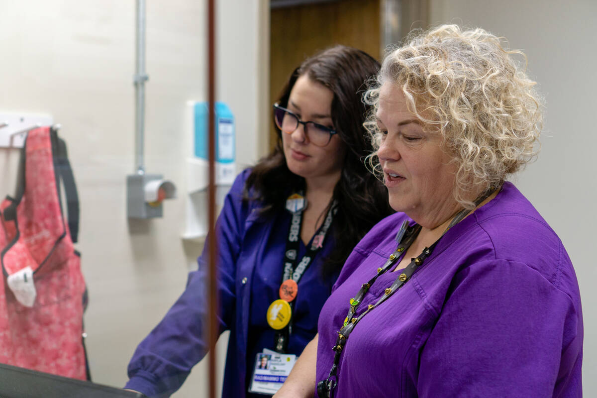
Angela Weiss has been working at Ivinson Memorial Hospital’s radiology department since 2002. She has spent 12 of those years in Ivinson’s Breast Imaging Center working in mammography.
As the Lead Mammographer, she has seen the mammogram process and equipment evolve over time. One thing she wants you to know, we are a long way from those horror stories you have heard about mammograms.
“We are hearing from our older generations about when mammograms were intimidating and very uncomfortable and that is what is being told to the next generation,” Angela said. “We need to get rid of those misconceptions.”
When Angela first joined Ivinson’s radiology department, imaging was still done on film. The timely process demanded more man power, longer wait times and lesser quality images.
“They had big machines that were called film rollers that they would hang films up for the radiologist and fold film,” Angela said. “It was a lot of footwork.”
Leaving films and the dark room as a thing of the past, Ivinson upgraded to a digital 2D machine that decreased wait times for patients and allowed radiologists to review images even faster.
In 2017, Ivinson’s Breast Imaging Center went even further to improve mammograms for patients by investing in the Hologic 2D/3D mammography machine. The advanced technology gives radiologists the tools to help identify breast cancer sooner, while providing a more comfortable and efficient patient experience.
“At Ivinson, we have a state-of-the-art Breast Imaging Center and our equipment is the best on the market. I have patients that have historically gone down south and they come here now because our equipment is top of the line,” Angela said.
The mammogram technicians at Ivinson are all licensed and frequently trained on new education and positioning techniques that provide the best imaging and most comfort to a patient. Mammograms performed on the Hologic 2D/3D mammogram machine use a variety of paddles that provide better breast compression while using less force. Studies have found that 3D mammograms offer better evaluations for patients with dense breast tissue.
“It’s more comfortable for the patient and it also gives us a better read-out because we have a better consistency of compression throughout the whole breast. We also have a foam pad that we can use if you have sensitive skin or are prone to tears.”
In addition to comfort, Ivinson’s advanced mammogram machinery provides radiologists with enhanced images to review.
“Moving from the digital machine to the 3D machine, the clarity is night and day compared to now. Then, it was very cloudy white looking and now our images are very crisp and clean looking. We can see more details in the breast than ever before. With the 3D we are able to see 1 millimeter thin slices.”
What’s the big deal with 3D? To a radiologist and the patients they see, it’s everything.
“Patients used to get call backs because of overlapping tissue. The radiologist wouldn’t always be able to differentiate if there was overlap or if there was something there. Patients would have to come back for additional images,” Angela explains of the old process. “Now, the radiologist can actually scroll through the one millimeter thin slices and they can see if it is tissue laying on top of tissue or if it is something laying within the tissue that we need to be concerned about. The resolution of the pictures is so good now, that we are picking up microcalcifications that we would have never seen before.”
Breast calcifications are deposits of calcium that develop in breast tissue. Calcifications are categorized as microcalcifications or macrocalcifications. Microcalcifications appear on mammograms as small white specks and can be a precursor for cancer.
“They are not always cancerous,” Angela explains. “We can have calcifications that are very benign in our breast but they can be cancerous and those cannot be picked up by any other modalities. That is why a mammogram is the gold standard. We are looking for calcifications and those are never going to be seen in ultrasounds.”
Most clinical guidelines recommend that women of average risk begin breast cancer screenings at age 40 with an annual mammogram. Patients with higher risks may need to be screened earlier and more often. Screening mammograms are routine and look for signs of breast cancer in patients that are not displaying signs or symptoms. Patients that are experiencing symptoms should not delay in contacting their healthcare provider.
“Get your mammogram,” Angela stresses. “I did not have my own until I worked as a mammographer and what made me get a mammogram was seeing what I do here at work. If I didn’t work in this field, I don’t know if I would have done it yet, because I didn’t really know the importance of it. Now, I cannot stress it enough.”
Like many women, Angela considered herself healthy, she had no family history of breast cancer and was considered at average risk. In the United States, one in eight women will develop invasive breast cancer in their lifetime and 85% of breast cancers have no family history at all.
As an expert in her field, Angela stresses that there is no good reason not to get your mammogram.
“98% of the young women that come for their first mammogram, walk out my door and say, this was nothing, and they are super relieved,” Angela said. After a long day of breast cancer screenings, her work does not stop there. Angela works to encourage awareness with not only her patients, but their inner circles as well. “I tell them, you know what your job is? Your job is to tell your friends and tell their friends, because breast cancer can happen to anybody.”
To schedule your yearly mammogram, ask your healthcare provider if they have standing mammogram orders at Ivinson’s Breast Imaging Center. Schedule your screening today at (307) 755‑4640.